Preventing and Managing Pressure Ulcers: A Comprehensive Guide
Pressure ulcers are among the most challenging complications in healthcare today, affecting millions of patients in hospitals, nursing homes, and home care settings each year. These painful wounds, also known as bedsores or decubitus ulcers, can develop in as little as two hours and significantly impact quality of life, mobility, and overall health. At Healogics, we understand the physical and emotional toll that pressure sores take on patients and their loved ones. Our mission, FIND. TREAT. HEAL.™ drives us to provide the most advanced, compassionate care possible while empowering patients and caregivers with the knowledge they need to prevent and manage these serious wounds.
The statistics surrounding pressure ulceration are sobering. Research indicates that approximately 2.5 million people in the United States develop pressure ulcers annually, with rising prevalence in hospitals and long-term care facilities. Beyond physical suffering, these wounds place an enormous burden on the healthcare system. More importantly, pressure ulcers can lead to severe complications, extended hospital stays, and reduced independence for those affected. We believe that through proper education, prevention strategies, and timely intervention, many of these wounds can be avoided or successfully treated before they progress to more serious stages.
Pressure Ulcers: An Overview
A pressure ulcer is a localized injury to the skin and underlying tissue that results from sustained pressure, friction, or shear forces on the body. When pressure restricts blood flow to vulnerable areas for extended periods, the affected tissue becomes deprived of oxygen and essential nutrients, leading to cellular death and wound formation. We often see these injuries referred to by several names, including bedsores, pressure sores, and decubitus ulcers, all describing the same condition that develops when skin and tissue are compressed against bone for too long.
Understanding the nature of pressure ulcers is the first step in preventing them. Unlike other types of wounds that result from external trauma, pressure ulcers develop from the inside out. The damage often begins in the deeper tissue layers near the bone before becoming visible on the skin’s surface. This characteristic makes early detection particularly challenging and underscores the importance of regular skin assessments for at-risk individuals.
What Causes Bed Sores?
The development of bed sores stems from three primary mechanical forces that compromise skin integrity and tissue health. We want to ensure patients and caregivers understand these causes to better prevent pressure ulceration:
Sustained Pressure
The most significant factor in developing a pressure ulcer is prolonged, unrelieved pressure on the skin. When body weight presses the skin against a firm surface, such as a mattress or chair, blood vessels are compressed, reducing or completely blocking blood flow to the area. Without adequate circulation, tissue cells are deprived of oxygen and nutrients, leading to tissue breakdown. This process can begin in as little as two to three hours in vulnerable individuals.
Friction
Friction occurs when skin rubs against clothing, bedding, or other surfaces. This repetitive rubbing can remove the skin’s protective outer layer, making the area more susceptible to injury. We commonly observe friction-related damage in patients who slide down in their chair or bed, or who are moved without proper lifting techniques.
Shear Forces
Shear represents one of the most damaging forces contributing to pressure sore development. It occurs when layers of skin move in opposite directions, such as when a patient’s body slides down while their skin remains stuck to the bed surface. This internal stretching and tearing of tissue and blood vessels can cause significant damage to deeper tissue layers, even when the skin surface appears intact.
Additional contributing factors that increase the risk of decubitus ulcer formation include moisture from perspiration, urine, or wound drainage, which can weaken skin and make it more vulnerable to breakdown. Poor circulation, particularly in older adults or those with vascular diseases, further compromises the skin’s ability to withstand pressure.
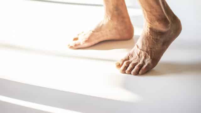
What Are the Common Sites?
Pressure ulcers typically develop over bony prominences where there is less natural cushioning between the bone and skin surface. We want patients and caregivers to pay special attention to these high-risk areas:
- Sacrum and Tailbone: The most common site for pressure sores in bedridden patients, accounting for approximately 30% to 40% of all pressure ulcers
- Heels: Particularly vulnerable in patients lying in bed, with limited ability to change position
- Hips and Sides of the Pelvis: Common sites for those who lie on their sides for extended periods
- Shoulder Blades: Often affected in patients who spend significant time in a semi-reclined position
- Back of the Head: Especially in infants and older adults who cannot reposition themselves
- Elbows: Vulnerable patients who rest on their elbows while in bed
- Ankles: Can develop pressure ulcers when legs remain in one position for too long
- Buttocks: Particularly at risk for those who spend extended time sitting in a chair or wheelchair
The specific location where a pressure ulcer develops often depends on the patient’s typical positioning. Those who are primarily bedridden tend to develop wounds on the sacrum, heels, and back of the head, while individuals who spend most of their time in a wheelchair or chair are more likely to develop pressure sores on the buttocks, tailbone, shoulder blades, and backs of the arms and legs.
Who is at Risk of Pressure Ulcers?
Understanding risk factors helps us identify individuals who need enhanced preventive care. We’ve identified several key factors that significantly increase the likelihood of developing a decubitus ulcer:
Immobility and Limited Movement
Individuals who cannot change positions without assistance face the highest risk. This includes those who are bedridden, paralyzed, sedated, in a coma, or recovering from surgery. Even patients who can move but do so infrequently are at elevated risk.
Advanced Age
Older adults are particularly vulnerable to pressure ulcers for several reasons. As we age, skin becomes thinner, less elastic, and more fragile. Older individuals may also have reduced sensation, making them less aware of discomfort that would normally prompt repositioning. Additionally, older adults often have multiple chronic conditions that impair healing.
Nutritional Deficiencies
Inadequate nutrition and dehydration significantly impair the skin’s ability to maintain its integrity and repair damage. Protein deficiency, in particular, compromises tissue strength and healing capacity. Patients who have difficulty eating or drinking independently, or those with conditions that affect nutrient absorption, face a higher risk.
Chronic Medical Conditions
Several health conditions increase susceptibility to pressure ulceration:
- Diabetes, which can reduce sensation and impair circulation and wound healing
- Vascular diseases that compromise blood flow to the skin
- Heart disease that reduces overall circulation
- Kidney disease that can lead to fluid retention and skin fragility
- Neurological conditions like spinal cord injuries, multiple sclerosis, or stroke that reduce sensation and mobility
- Dementia or confusion that may prevent individuals from recognizing or responding to discomfort
Moisture and Incontinence
Prolonged exposure to urine, feces, or excessive perspiration breaks down the skin’s protective barrier, making it more vulnerable to pressure damage. Moisture also increases friction between skin and surfaces.
Previous Pressure Ulcer
Individuals who have had a pressure sore before are at higher risk of developing new ones, as previously damaged skin may have reduced strength and resilience.
Poor Circulation and Reduced Sensation
Conditions that impair blood flow or nerve function make it difficult for the body to deliver adequate oxygen and nutrients to at-risk areas. Reduced sensation also means patients may not feel the discomfort that would typically trigger repositioning.
How to Identify and Diagnose Bed Sores
Early identification of pressure ulcers is crucial for successful treatment and prevention of progression to more severe stages. We emphasize the importance of regular, thorough skin assessments for all at-risk individuals. Daily inspection of vulnerable areas, particularly bony prominences, should be part of routine care for anyone with limited mobility.
When we assess patients for pressure ulcer risk, we often use standardized tools like the Braden Scale, which evaluates six key factors: sensory perception, moisture exposure, activity level, mobility, nutritional status, and friction/shear forces. This assessment helps us identify those who need enhanced preventive interventions and allows us to implement targeted strategies before tissue damage occurs.
Diagnosing a pressure ulcer involves careful visual inspection and gentle palpation of the skin. We look for changes in skin color, temperature, texture, firmness, and the presence of any breaks in the skin. It’s important to note that in individuals with darker skin tones, pressure ulcers may not present as obvious redness. Instead, we watch for areas that appear darker, purplish, or bluish compared to surrounding skin, as well as changes in skin temperature or firmness.
Stages and Symptoms of Pressure Ulcers
Understanding pressure ulcer stages is essential for determining appropriate treatment and monitoring healing progress. We classify pressure sores into four distinct stages based on the depth and severity of tissue damage, along with two additional categories for wounds that cannot be fully assessed:
Stage 1 Pressure Ulcer
At this earliest stage, the skin remains intact but shows signs of damage. The area appears red or discolored and does not blanch (turn white) when pressed. In darker skin tones, the area may appear darker than the surrounding tissue or have a purple or blue hue. The affected area may feel warmer or cooler than adjacent tissue and may be firm or soft. Patients often report pain, tingling, or itching at the site. Stage 1 pressure ulcers are reversible with prompt intervention and pressure relief.
Stage 2 Pressure Ulcer
At this stage, the skin breaks open, and the wound appears as a shallow, open ulcer with a pink or red wound bed. It may present as an intact or ruptured blister filled with clear fluid or blood. The wound affects the epidermis (the outer skin layer) and may extend into the dermis (the deeper skin layer). The area is typically painful, and the surrounding tissue may show signs of damage. With proper care, Stage 2 pressure sores can heal within weeks to months.
Stage 3 Pressure Ulcer
These deeper wounds extend through the full thickness of the skin into the subcutaneous fat layer. The ulcer appears as a crater-like wound, and yellow or tan dead tissue (slough) or black dead tissue (eschar) may be present. The wound base is typically red or pink and may show signs of undermining or tunneling, where the wound extends beneath intact skin at the edges. Bone, tendon, and muscle are not visible at this stage, but the wound is deep enough that fat may be exposed. Stage 3 pressure ulcers require professional wound care and can take months to heal.
Stage 4 Pressure Ulcer
The most severe category, Stage 4 pressure ulcers involve extensive tissue destruction with damage extending through skin, fat, and muscle, potentially exposing bone, tendon, or joint. These deep wounds often contain slough or eschar and frequently exhibit undermining and tunneling. Complications such as osteomyelitis (bone infection) are common. Stage 4 pressure sores require intensive medical treatment and may take months to years to heal, sometimes requiring surgical intervention.
Unstageable Pressure Ulcer
When the wound base is covered by yellow, tan, gray, green, or brown slough, or by tan, brown, or black eschar, we cannot accurately assess the depth of tissue damage. These wounds are classified as unstageable until enough dead tissue is removed to expose the wound base and determine the true stage.
Deep Tissue Pressure Injury
This category describes areas where the skin appears intact but shows a purple or maroon discoloration or appears as a blood-filled blister. These injuries indicate damage to underlying soft tissue from pressure or shear forces. The area may be painful, firm, mushy, or boggy compared to adjacent tissue. Deep tissue injuries are particularly concerning because significant damage may exist beneath apparently intact skin, and these wounds can rapidly evolve into Stage 3 or 4 ulcers.
The pressure ulcer symptoms patients experience vary by stage but commonly include pain or discomfort at the site, changes in skin color or temperature, swelling, drainage from the wound, and, in advanced stages, foul odor indicating infected tissue. It’s crucial to monitor any pressure sore for signs of infection, including increased pain, redness, warmth, swelling, pus or cloudy drainage, fever, or a foul smell from the wound.
Pressure Sore Treatment and Management
Effective pressure ulcer treatment requires a comprehensive, individualized approach that addresses both the wound itself and the underlying factors that contributed to its development. We believe in treating the whole person, not just the wound, which is why our care plans consider all aspects of a patient’s health and circumstances.
The foundation of pressure ulcer management involves relieving pressure on the affected area, keeping the wound clean, managing pain, preventing infection, and optimizing overall health to support healing. The specific treatments we recommend depend on the stage of the pressure ulcer, its location, the presence of infection, and the patient’s overall health status.
Wound Care
Proper wound care forms the cornerstone of pressure ulcer treatment. We begin by carefully cleaning the wound with gentle, non-toxic solutions, typically normal saline or clean water, to remove debris and bacteria without damaging healing tissue. Harsh antiseptics like hydrogen peroxide or iodine should be avoided, as they can harm healthy cells and delay healing.
After cleaning, we apply appropriate dressings that maintain a moist wound environment, which research has shown promotes faster healing compared to dry wound care. The type of dressing we select depends on the pressure ulcer stage, amount of drainage, and presence of dead tissue:
For Stage 1 pressure ulcers, transparent film dressings or thin hydrocolloid dressings protect the area while allowing us to monitor the skin. For Stage 2 wounds, hydrocolloid dressings or foam dressings absorb drainage while maintaining moisture balance. Stage 3 and 4 pressure ulcers often require more advanced dressings such as alginates for heavily draining wounds, hydrogels to maintain moisture in drier wounds, or specialized foam dressings that can manage exudate while providing cushioning.
The frequency of dressing changes varies depending on the product used and the amount of wound drainage, but we typically recommend changes every 1 to 7 days. It’s essential to monitor the wound at each dressing change for signs of healing or complications. We teach patients and caregivers to watch for signs of infection, increased pain, changes in drainage color or odor, and any expansion of the wound.
One critical aspect of wound care is protecting the surrounding skin. We apply barrier products to prevent moisture from breaking down healthy skin adjacent to the wound, which could enlarge the pressure sore. Proper wound care not only promotes healing but also prevents complications that could worsen the patient’s condition.
Pain Management
We recognize that pressure ulcers can be extremely painful, and managing this pain is essential for patient comfort and quality of life. Pain can interfere with sleep, mobility, and overall well-being, so we take a multifaceted approach to pain relief.
For mild to moderate pain, over-the-counter medications like acetaminophen may provide relief. More severe pain may require prescription pain medications, including opioids in some cases. We work closely with patients to find the most effective pain management regimen with the fewest side effects.
Topical treatments, such as lidocaine-based creams or gels, can provide localized pain relief, particularly during dressing changes. Some patients benefit from applying these agents before wound care procedures to reduce discomfort.
Beyond medications, we recommend several complementary approaches to pain management. Ensuring proper positioning with adequate cushioning can significantly reduce pressure-related pain. Relaxation techniques, including deep breathing exercises, guided imagery, and meditation, help some patients cope with chronic pain. Some individuals find relief through gentle massage of surrounding areas (never directly on the wound), warm compresses applied to nearby regions, or distraction techniques during painful procedures.
It’s important to note that increased pain can signal complications such as infection or wound deterioration, so we encourage patients to report any changes in pain levels to their healthcare team immediately.
Antibiotics
Not all pressure ulcers require antibiotic treatment. We reserve antibiotics for cases where infection is present or there is a significant risk of systemic infection developing. Determining when antibiotics are necessary requires careful assessment by healthcare professionals.
Signs that a pressure ulcer may be infected include increased redness, warmth, or swelling around the wound; increased pain or tenderness; purulent (pus-like) drainage that may be yellow, green, or gray; foul odor from the wound; fever or chills; and increased wound size or depth despite appropriate treatment. In severe cases, infection can spread beyond the wound, leading to cellulitis (infection of the surrounding tissue) or even sepsis (a life-threatening bloodstream infection).
When infection is confirmed through clinical assessment or wound cultures, we prescribe appropriate antibiotics based on the specific bacteria involved. Oral antibiotics may suffice for localized infections, whereas more severe or systemic infections require intravenous antibiotics, which may necessitate hospitalization.
It’s crucial to complete the entire course of prescribed antibiotics, even if the wound appears to be improving, to prevent antibiotic resistance and ensure complete eradication of the infection. We also emphasize that antibiotics alone cannot heal a pressure ulcer. They must be combined with appropriate wound care, pressure relief, and treatment of underlying factors.
Debridement
Debridement is the medical process of removing dead, damaged, or infected tissue from a wound to promote healing. Dead tissue, whether it appears as yellow slough or black eschar, provides a breeding ground for bacteria and prevents the wound from healing. We cannot fully assess the depth of a pressure ulcer until this tissue is removed, which is why debridement is often one of the first steps in treating more advanced pressure sores.
We have several debridement methods available, and we select the most appropriate technique based on the wound characteristics, the patient’s overall health, and the urgency of the situation:
Surgical Debridement
This method uses surgical instruments to remove large amounts of dead tissue quickly. It’s the fastest debridement technique and is typically reserved for urgent situations, such as when infection is spreading rapidly or when large amounts of dead tissue are present. Surgical debridement may be performed at the bedside for limited areas or in an operating room for more extensive procedures.
Mechanical Debridement
These techniques physically remove dead tissue using methods such as wet-to-dry dressings (now less commonly used due to potential damage to healthy tissue) or wound irrigation with pressurized saline. Mechanical debridement is non-selective, meaning it can remove both dead and healthy tissue, so we use it carefully.
Enzymatic Debridement
This approach uses topical medications containing enzymes that break down dead tissue. We apply these enzymatic debriding agents directly to the wound, and they work gradually over several days to weeks. This method is useful for patients who cannot tolerate surgical debridement or for those with dead tissue firmly attached.
Autolytic Debridement
The body’s own enzymes and moisture can naturally break down dead tissue. We facilitate this process by using moisture-retentive dressings that create an optimal environment for the body’s natural debridement mechanisms. This is the slowest but most selective method, affecting only dead tissue. It’s appropriate for wounds without infection and when there’s no urgency to remove dead tissue quickly.
The choice of debridement method depends on multiple factors, and sometimes we combine techniques for optimal results. Regular assessment during debridement ensures we remove all non-viable tissue while preserving healthy tissue that’s essential for healing.
Surgery
While most pressure ulcers heal with conservative treatment, some severe cases require surgical intervention. We typically consider surgery for Stage 3 or 4 pressure ulcers that haven’t responded to other treatments, large wounds that would take an extremely long time to heal on their own, or recurrent pressure sores at the same site.
Surgical options for pressure ulcer treatment include:
Pressure Ulcer Closure
For some deep wounds, surgeons can thoroughly clean the wound, remove dead tissue, and close it with sutures. This approach is only suitable for certain wounds and requires careful patient selection.
Flap Surgery
This is the most common surgical procedure for severe pressure ulcers. The surgeon uses tissue from adjacent areas (muscle, skin, or both) to cover the wound and pad the bony prominence. These tissue flaps bring their own blood supply, which promotes healing and provides cushioning to prevent future ulcers. Flap surgery is a major procedure requiring general anesthesia and significant recovery time.
Skin Grafts
In some cases, surgeons harvest skin from another area of the patient’s body and use it to cover the pressure ulcer. Skin grafts are typically thinner than flaps and don’t provide the same cushioning, but they can be appropriate for certain wounds.
Surgery for pressure ulcers comes with significant risks, including infection, bleeding, anesthesia complications, and failure of the surgical repair. Recovery can take weeks to months, and patients must strictly adhere to pressure-relief protocols afterward, as the repaired area is at high risk of developing new pressure sores. Success rates vary, but recurrence of pressure ulcers after surgical repair ranges from 20% to 30%, making prevention strategies essential even after surgery.
We carefully evaluate each patient’s overall health, nutritional status, and ability to comply with post-surgical care requirements before recommending surgical treatment. In many cases, optimizing conservative treatment approaches yields better outcomes than surgery.
Other Treatments
In addition to standard pressure sore treatment approaches, we offer several advanced therapies that can enhance healing in difficult-to-treat wounds:
Negative Pressure Wound Therapy (NPWT)
This treatment uses a specialized dressing connected to a vacuum pump that applies controlled negative pressure to the wound. NPWT removes excess fluid, promotes blood flow, draws wound edges together, and stimulates the formation of granulation tissue. We often use this therapy for Stage 3 or 4 pressure ulcers with significant depth or drainage.
Hyperbaric Oxygen Therapy
This advanced treatment involves breathing pure oxygen in a pressurized chamber, which significantly increases the amount of oxygen in the blood and tissues. Enhanced oxygen delivery promotes wound healing by supporting cellular repair processes, combating certain bacteria, and reducing swelling. Hyperbaric oxygen therapy can be particularly beneficial for pressure ulcers complicated by infection or poor circulation.
Growth Factors and Biological Dressings
These advanced products contain substances that stimulate cellular growth and tissue repair. Platelet-derived growth factors, for example, can accelerate healing in chronic wounds. Biological dressings made from materials like collagen or other natural substances provide a scaffold for new tissue growth.
At Healogics-managed Wound Care Centers®, many of these advanced therapies are offered as part of comprehensive treatment plans tailored to each patient’s specific needs. Our wound care specialists stay current with the latest research and technologies to provide the most effective treatments available.
How to Prevent Pressure Ulcers
Prevention is always preferable to treatment for pressure ulcers. We believe that with proper care, vigilance, and adherence to evidence-based strategies, most pressure sores can be prevented. The key to pressure ulcer prevention lies in eliminating or minimizing the mechanical forces that cause tissue damage while optimizing overall health to maintain skin integrity.
Repositioning and Mobility
The single most important strategy for preventing pressure ulcers is regular repositioning to relieve pressure on vulnerable areas. We recommend that bedridden patients change position at least every two hours, though some high-risk individuals may need more frequent repositioning. Those who can reposition themselves should be encouraged to do so even more frequently: shifting weight every 15 to 30 minutes if possible.
When repositioning patients in bed, we follow these best practices: use a 30-degree angle when turning patients on their sides, rather than a full 90-degree side-lying position, to reduce pressure on the hips; avoid positioning directly on bony prominences like the hips or tailbone; use pillows or foam wedges to keep knees and ankles from touching each other; elevate the head of the bed no more than 30 degrees to prevent sliding down (which causes shear forces); and keep heels completely off the bed surface using pillows under the calves.
For individuals who spend significant time in a chair or wheelchair, repositioning is equally important. We recommend that wheelchair users perform pressure-relief movements every 15 to 30 minutes by lifting themselves off the seat using the armrests, leaning forward, or leaning from side to side. Those who cannot reposition themselves should be assisted to change position at least every hour.
Encouraging mobility whenever possible is crucial. Even patients with limited mobility can often participate in range-of-motion exercises or transfer activities that reduce time spent in any single position. Physical therapy can help maintain or improve mobility, which indirectly reduces pressure ulcer risk.
Skin Care and Hygiene
Maintaining healthy, intact skin is essential for preventing pressure ulcers. We recommend a comprehensive skin care approach that includes daily inspection of all high-risk areas for redness, warmth, swelling, or changes in skin color or texture. For individuals with limited mobility, caregivers should perform thorough skin assessments during bathing and repositioning.
Proper cleansing is important, but should be gentle. We advise using mild, pH-balanced cleansers rather than harsh soaps that can dry out skin. Water temperature should be warm, not hot, as excessive heat can damage fragile skin. After bathing, pat skin dry rather than rubbing, paying special attention to skin folds where moisture can accumulate.
Moisturizers play a vital role in maintaining skin health. We recommend applying moisturizing lotions or creams at least once a day to prevent dry, cracked skin that’s more prone to breakdown. However, avoid applying moisturizer to areas of redness that might indicate the early stage of a pressure ulcer, as this can trap moisture against already compromised skin.
Managing moisture is equally important as preventing dryness. Excess moisture from perspiration, urine, or feces can macerate skin, making it soft and more susceptible to damage. For patients with incontinence, we recommend checking and changing briefs or pads frequently, using moisture-barrier creams or ointments to protect the skin, and considering a bowel or bladder management program to reduce exposure to moisture.
Avoid massaging over bony prominences or reddened areas. While massage might seem beneficial, research shows it can actually damage tissue that’s already compromised. Finally, minimize exposure to friction by using lifting techniques rather than dragging patients across surfaces, ensuring bed linens are smooth and wrinkle-free, and applying protective dressings or barrier films to areas prone to friction.
Nutrition and Hydration
Adequate nutrition is fundamental to maintaining skin integrity and supporting healing if pressure ulceration does occur. We work with patients and caregivers to ensure nutritional needs are met through a diet rich in essential nutrients:
Protein is particularly crucial, as it’s the building block for tissue repair and maintenance. We typically recommend 1.2 to 1.5 grams of protein per kilogram of body weight daily for at-risk individuals. Good protein sources include lean meats, fish, eggs, dairy products, legumes, and nuts.
Vitamins and minerals play specific roles in skin health. Vitamin C supports collagen formation and wound healing; vitamin A helps maintain skin integrity and supports immune function; vitamin E has antioxidant properties that protect cells; zinc is essential for protein synthesis and immune function; and B vitamins support overall skin health and energy metabolism.
Hydration is often overlooked but is critical for maintaining skin elasticity and preventing breakdown. We encourage adequate fluid intake, typically six to eight glasses of water daily unless medical conditions require fluid restriction. Dehydration makes skin dry, less elastic, and more prone to damage.
For patients who cannot meet nutritional needs through regular meals, we may recommend nutritional supplements, high-protein snacks between meals, or consultation with a registered dietitian to develop a comprehensive nutrition plan. In some cases, enteral nutrition (tube feeding) may be necessary to ensure adequate nutrient intake.
Maintaining a healthy body weight is also important, as both significant underweight and obesity increase the risk of pressure ulcers. Underweight individuals lack the natural cushioning that protects bony prominences, while excess weight increases pressure forces and can make repositioning more difficult.
Smoking Cessation
Smoking significantly impairs wound healing and increases pressure ulcer risk through multiple mechanisms. Nicotine causes blood vessels to constrict, reducing blood flow to the skin and limiting the delivery of oxygen and nutrients. Carbon monoxide from cigarette smoke binds to hemoglobin, further reducing oxygen availability to tissues. Smoking also impairs immune function, making the body less able to fight infection and repair damage.
We strongly encourage all patients at risk for pressure ulcers to quit smoking. While we understand that smoking cessation can be challenging, the benefits for wound prevention and healing are substantial. We can provide information about smoking cessation programs, nicotine replacement therapies, prescription medications that reduce cravings, and behavioral counseling to support patients in quitting.
Even reducing the number of cigarettes smoked daily can provide some benefit, though complete cessation offers the greatest improvement in tissue health and healing capacity.
Use of Support Surfaces
Specialized support surfaces, including mattresses, overlays, and cushions, are designed to redistribute pressure, reduce friction and shear, and control moisture. These devices are essential components of pressure ulcer prevention for at-risk individuals.
Support surface types include:
Foam Mattresses and Overlays
High-quality foam products with proper thickness (typically 3 to 4 inches or more) can effectively redistribute pressure. Some foam surfaces feature contoured or egg-crate designs that improve pressure distribution.
Air-Filled or Dynamic Support Surfaces
These devices use air cells that inflate and deflate in cycles, continuously changing pressure points. This “alternating pressure” technology is particularly beneficial for high-risk individuals. Some air mattresses allow for pressure adjustment based on patient weight and comfort.
Low-Air-Loss Beds
These specialized beds continuously blow air through small holes in the mattress surface, which helps control moisture and temperature while distributing pressure. They’re typically reserved for patients at very high risk or those who already have pressure ulcers.
Pressure-Relieving Cushions
For individuals who spend significant time in chairs or wheelchairs, specialized cushions made from foam, gel, air, or combinations of these materials help distribute pressure away from bony prominences. Wheelchair cushions should be selected based on the individual’s weight, posture, and activity level.
It’s important to note that no support surface eliminates the need for repositioning. Even the best mattress or cushion cannot completely prevent pressure ulcers without regular position changes. Additionally, standard hospital mattresses and regular home mattresses typically do not provide adequate pressure redistribution for at-risk individuals.
When selecting a support surface, consider the patient’s risk level, existing pressure ulcers, body size and weight, mobility level, and environmental factors such as heat and moisture. Proper maintenance of support surfaces, including keeping them clean and ensuring proper inflation of air surfaces, is essential to their effectiveness and safety.
Complications of Pressure Ulcers
When pressure ulcers are not properly treated or prevention strategies fail, serious complications can develop, significantly impacting health and quality of life. We want patients and caregivers to understand these potential complications to emphasize the importance of early intervention and comprehensive care.
Infection represents the most common and concerning complication. Pressure ulcers provide an entry point for bacteria, and the damaged tissue serves as a breeding ground for infection. Cellulitis, a spreading infection of the skin and soft tissues surrounding the wound, can develop and cause significant pain, swelling, and systemic illness. Without prompt antibiotic treatment, cellulitis can progress to more severe infections.
Sepsis, a life-threatening condition, occurs when infection spreads into the bloodstream, triggering a systemic inflammatory response. Sepsis can lead to organ failure and death if not recognized and treated immediately. Older adults and those with compromised immune systems face a particularly high risk. We teach caregivers to watch for warning signs, including fever or abnormally low body temperature, rapid heart rate, confusion or decreased alertness, and severe weakness.
Osteomyelitis is a bone infection that can occur when deep pressure ulcers extend to the bone. This serious complication requires prolonged antibiotic therapy, sometimes for weeks or months, and may necessitate surgical removal of infected bone. Osteomyelitis can be difficult to eradicate completely and may become chronic.
Joint and Tendon Damage can occur when Stage 3 or 4 pressure ulcers expose or extend into joints or tendons. This damage can severely limit mobility and function, potentially requiring surgical intervention and extensive rehabilitation.
Chronic Wounds may develop when pressure ulcers fail to heal despite appropriate treatment. These non-healing wounds can persist for months or years, causing ongoing pain, limiting mobility, reducing quality of life, and increasing the risk of other complications.
When to Seek Medical Help
When in doubt, it’s always safer to involve a professional. Seek medical help if a pressure ulcer shows signs of infection (such as redness, warmth, swelling, odor, or drainage), if pain increases, or if the wound fails to improve after a few days of proper care. Individuals with diabetes, poor circulation, limited mobility, or a history of slow-healing wounds should reach out even sooner. A healthcare provider can offer a thorough assessment and recommend a personalized treatment plan to prevent complications.
If you have concerns about a pressure ulcer, contact your local Wound Care Center® or request an appointment today.